
keyword
medical image segmentation
Medical image segmentation is the process of partitioning a digital medical scan into distinct, meaningful regions by assigning a specific anatomical or pathological label to each individual pixel or voxel. Operating on two-dimensional images or volumetric three-dimensional scans from modalities such as magnetic resonance imaging, computed tomography, ultrasound, endoscopy, and microscopy, this technique delineates the precise boundaries and morphologies of target structures, including organs, bones, blood vessels, cellular nuclei, and abnormalities like tumors or lesions. By transforming raw visual imaging data into quantitative masks, medical image segmentation serves as a critical prerequisite for computer-aided diagnosis, radiation therapy targeting, surgical planning, disease progression monitoring, and quantitative biomarker analysis, and is widely automated using advanced deep learning architectures designed to capture multi-scale spatial and semantic relationships.
21 items
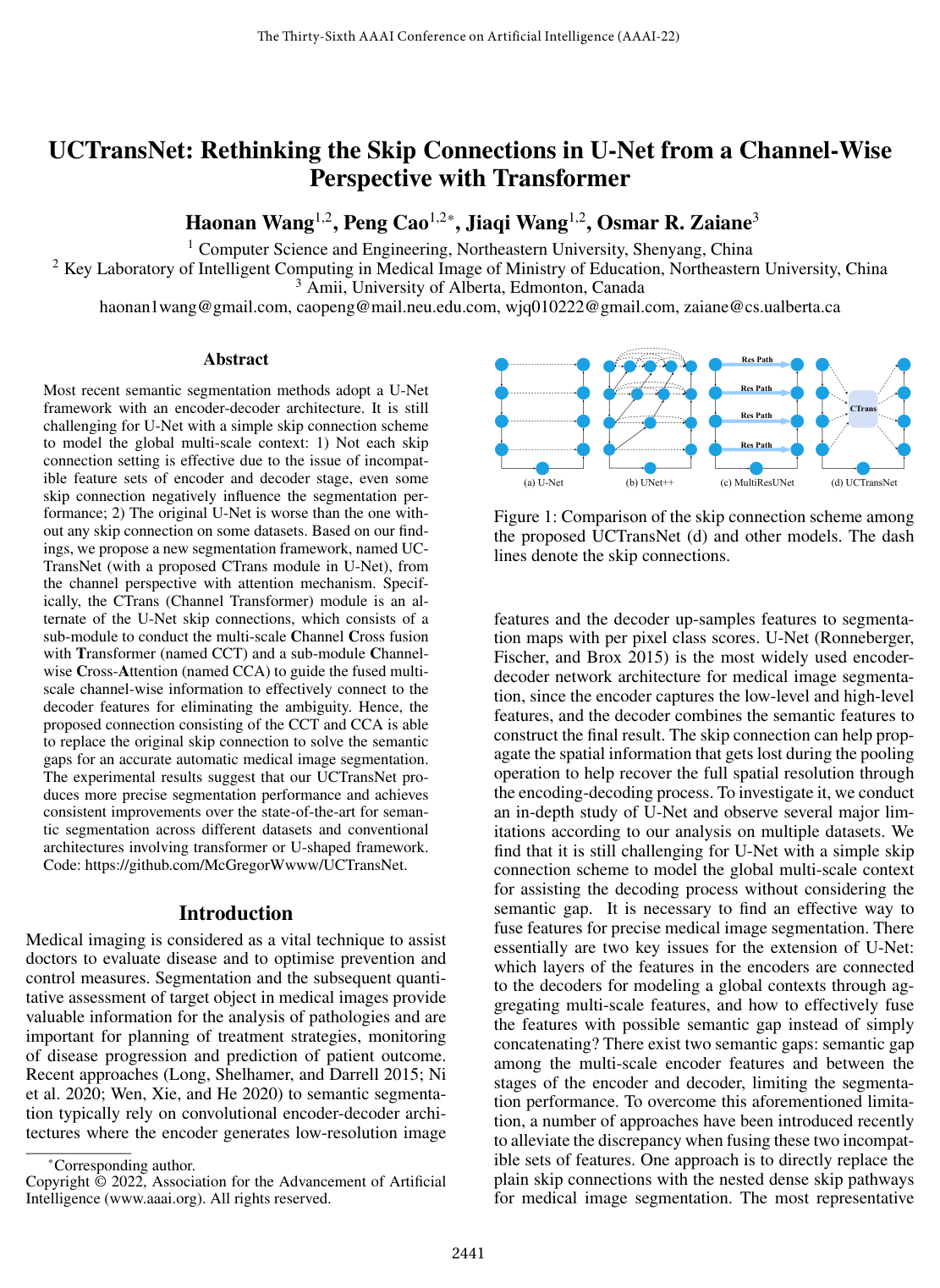
UCTransNet: Rethinking the Skip Connections in U-Net from a Channel-Wise Perspective with Transformer
Haonan Wang, Peng Cao, Jiaqi Wang, Osmar R. Zaïane
Why you should read this
Proposes a channel-wise transformer module that replaces traditional U-Net skip connections to bridge semantic gaps between encoder and decoder features, consistently improving medical image segmentation accuracy across multiple benchmarks.
Most recent semantic segmentation methods adopt a U-Net framework with an encoder-decoder architecture. It is still challenging for U-Net with a simple skip connection scheme to model the global multi-scale context: 1) Not each skip connection setting is effective due to the issue of incompatible feature sets of encoder and decoder stage, even some skip connection negatively influence the segmentation performance; 2) The original U-Net is worse than the one without any skip connection on some datasets. Based on our findings, we propose a new segmentation framework, named UC-TransNet (with a proposed CTrans module in U-Net), from the channel perspective with attention mechanism. Specifically, the CTrans (Channel Transformer) module is an alternate of the U-Net skip connections, which consists of a sub-module to conduct the multi-scale Channel Cross fusion with Transformer (named CCT) and a sub-module Channel-wise Cross-Attention (named CCA) to guide the fused multi-scale channel-wise information to effectively connect to the decoder features for eliminating the ambiguity. Hence, the proposed connection consisting of the CCT and CCA is able to replace the original skip connection to solve the semantic gaps for an accurate automatic medical image segmentation. The experimental results suggest that our UCTransNet produces more precise segmentation performance and achieves consistent improvements over the state-of-the-art for semantic segmentation across different datasets and conventional architectures involving transformer or U-shaped framework.
Added
2026-10-04
Anatomically-aware conformal prediction for medical image segmentation with random walks
Melanie Gaillochet Christian Desrosiers Hervé Lombaert, Melanie Gaillochet, Christian Desrosiers, Hervé Lombaert
Why you should read this
Introduces Random-Walk Conformal Prediction, a model-agnostic framework that diffuses uncertainty over vision foundation model feature graphs to generate spatially coherent, anatomically realistic segmentation prediction sets with strict statistical coverage guarantees.
The reliable deployment of deep learning in medical imaging requires uncertainty quantification that provides rigorous error guarantees while remaining anatomically meaningful. Conformal prediction (CP) is a powerful distribution-free framework for constructing statistically valid prediction intervals. However, standard applications in segmentation often ignore anatomical context, resulting in fragmented, spatially incoherent, and over-segmented prediction sets that limit clinical utility. To bridge this gap, this paper proposes Random-Walk Conformal Prediction (RW-CP), a model-agnostic framework which can be added on top of any segmentation method. RW-CP enforces spatial coherence to generate anatomically valid sets. Our method constructs a k-nearest neighbour graph from pre-trained vision foundation model features and applies a random walk to diffuse uncertainty. The random walk diffusion regularizes the non-conformity scores, making the prediction sets less sensitive to the conformal calibration parameter , ensuring more stable and continuous anatomical boundaries. RW-CP maintains rigorous marginal coverage while significantly improving segmentation quality. Evaluations on multi-modal public datasets show improvements of up to compared to standard CP baselines, given an allowable error rate of .
Added
2026-10-04
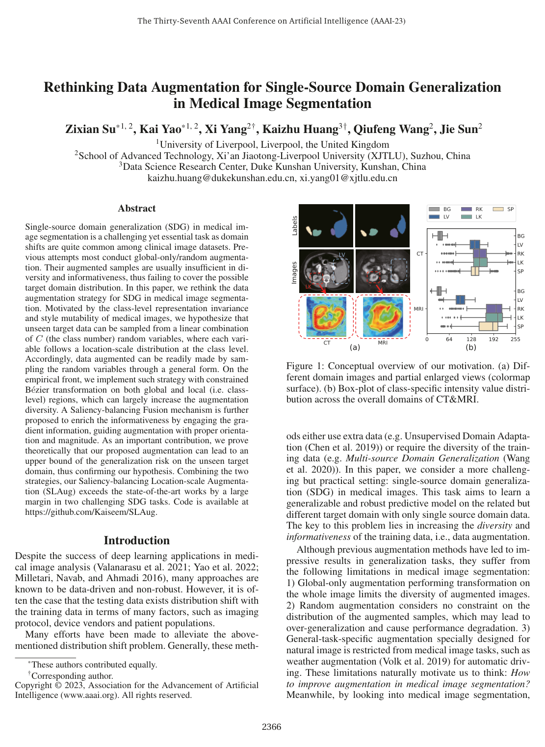
Rethinking Data Augmentation for Single-Source Domain Generalization in Medical Image Segmentation
Zixian Su, Kai Yao, Xi Yang, Kaizhu Huang, Qiufeng Wang, Jie Sun
Why you should read this
Proposes a class-level location-scale data augmentation framework paired with gradient-guided saliency balancing to guarantee bounded generalization risk and improve medical image segmentation across unseen domains.
Single-source domain generalization (SDG) in medical image segmentation is a challenging yet essential task as domain shifts are quite common among clinical image datasets. Previous attempts most conduct global-only/random augmentation. Their augmented samples are usually insufficient in diversity and informativeness, thus failing to cover the possible target domain distribution. In this paper, we rethink the data augmentation strategy for SDG in medical image segmentation. Motivated by the class-level representation invariance and style mutability of medical images, we hypothesize that unseen target data can be sampled from a linear combination of C (the class number) random variables, where each variable follows a location-scale distribution at the class level. Accordingly, data augmented can be readily made by sampling the random variables through a general form. On the empirical front, we implement such strategy with constrained Bézier transformation on both global and local (i.e. class-level) regions, which can largely increase the augmentation diversity. A Saliency-balancing Fusion mechanism is further proposed to enrich the informativeness by engaging the gradient information, guiding augmentation with proper orientation and magnitude. As an important contribution, we prove theoretically that our proposed augmentation can lead to an upper bound of the generalization risk on the unseen target domain, thus confirming our hypothesis. Combining the two strategies, our Saliency-balancing Location-scale Augmentation (SLAug) exceeds the state-of-the-art works by a large margin in two challenging SDG tasks. Code is available at https://github.com/Kaiseem/SLAug.
Added
2026-09-26
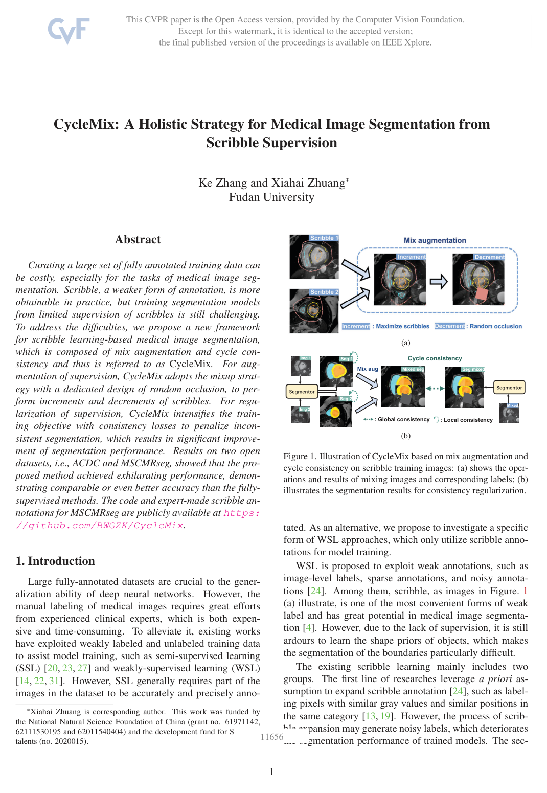
CycleMix: A Holistic Strategy for Medical Image Segmentation from Scribble Supervision
Ke Zhang, Xiahai Zhuang
Why you should read this
Proposes a weakly-supervised medical image segmentation framework that combines mixup-based scribble augmentation with global and local cycle-consistency regularization to achieve segmentation accuracy comparable to fully-supervised methods.
Curating a large set of fully annotated training data can be costly, especially for the tasks of medical image segmentation. Scribble, a weaker form of annotation, is more obtainable in practice, but training segmentation models from limited supervision of scribbles is still challenging. To address the difficulties, we propose a new framework for scribble learning-based medical image segmentation, which is composed of mix augmentation and cycle consistency and thus is referred to as CycleMix. For augmentation of supervision, CycleMix adopts the mixup strategy with a dedicated design of random occlusion, to perform increments and decrements of scribbles. For regularization of supervision, CycleMix intensifies the training objective with consistency losses to penalize inconsistent segmentation, which results in significant improvement of segmentation performance. Results on two open datasets, i.e., ACDC and MSCMRseg, showed that the proposed method achieved exhilarating performance, demonstrating comparable or even better accuracy than the fully-supervised methods. The code and expert-made scribble annotations for MSCMRseg are publicly available at https://github.com/BWGZK/CycleMix.
Added
2026-09-26
Class-Aware Adversarial Transformers for Medical Image Segmentation
Chenyu You, Ruihan Zhao, Fenglin Liu, Siyuan Dong, Sandeep Chinchali, Ufuk Topcu, Lawrence H. Staib, James S. Duncan
Why you should read this
Proposes CASTformer, a medical image segmentation framework combining multi-scale pyramid representations, a class-aware transformer module, and adversarial training to overcome standard tokenization limitations and substantially improve segmentation accuracy across multiple benchmarks.
Transformers have made remarkable progress towards modeling long-range dependencies within the medical image analysis domain. However, current transformer-based models suffer from several disadvantages: (1) existing methods fail to capture the important features of the images due to the naive tokenization scheme; (2) the models suffer from information loss because they only consider single-scale feature representations; and (3) the segmentation label maps generated by the models are not accurate enough without considering rich semantic contexts and anatomical textures. In this work, we present CASTformer, a novel type of adversarial transformers, for 2D medical image segmentation. First, we take advantage of the pyramid structure to construct multi-scale representations and handle multi-scale variations. We then design a novel class-aware transformer module to better learn the discriminative regions of objects with semantic structures. Lastly, we utilize an adversarial training strategy that boosts segmentation accuracy and correspondingly allows a transformer-based discriminator to capture high-level semantically correlated contents and low-level anatomical features. Our experiments demonstrate that CASTformer dramatically outperforms previous state-of-the-art transformer-based approaches on three benchmarks, obtaining 2.54%-5.88% absolute improvements in Dice over previous models. Further qualitative experiments provide a more detailed picture of the model's inner workings, shed light on the challenges in improved transparency, and demonstrate that transfer learning can greatly improve performance and reduce the size of medical image datasets in training, making CASTformer a strong starting point for downstream medical image analysis tasks.
Added
2026-09-26
Cross-patch Dense Contrastive Learning for Semi-supervised Segmentation of Cellular Nuclei in Histopathologic Images
Huisi Wu, Zhaoze Wang, Youyi Song, Lin Yang, Jing Qin
Why you should read this
Proposes a cross-patch dense contrastive learning framework that pairs patch- and pixel-level representations within a mean-teacher architecture to segment cellular nuclei accurately from limited annotated histopathology images.
We study the semi-supervised learning problem, using a few labeled data and a large amount of unlabeled data to train the network, by developing a cross-patch dense contrastive learning framework, to segment cellular nuclei in histopathologic images. This task is motivated by the expensive burden on collecting labeled data for histopathologic image segmentation tasks. The key idea of our method is to align features of teacher and student networks, sampled from cross-image in both patch- and pixel-levels, for enforcing the intra-class compactness and inter-class separability of features that as we shown is helpful for extracting valuable knowledge from unlabeled data. We also design a novel optimization framework that combines consistency regularization and entropy minimization techniques, showing good property in eviction of gradient vanishing. We assess the proposed method on two publicly available datasets, and obtain positive results on extensive experiments, outperforming the state-of-the-art methods. Codes are available at https://github.com/zzw-szu/CDCL.
Added
2026-09-26
Directional Connectivity-based Segmentation of Medical Images
Ziyun Yang, Sina Farsiu
Anatomical consistency in biomarker segmentation is crucial for many medical image analysis tasks. A promising paradigm for achieving anatomically consistent segmentation via deep networks is incorporating pixel connectivity, a basic concept in digital topology, to model inter-pixel relationships. However, previous works on connectivity modeling have ignored the rich channel-wise directional information in the latent space. In this work, we demonstrate that effective disentanglement of directional sub-space from the shared latent space can significantly enhance the feature representation in the connectivity-based network. To this end, we propose a directional connectivity modeling scheme for segmentation that decouples, tracks, and utilizes the directional information across the network. Experiments on various public medical image segmentation benchmarks show the effectiveness of our model as compared to the state-of-the-art methods. Code is available at https://github.com/Zyun-Y/DconnNet.
Added
2026-09-26
Closing the Generalization Gap of Cross-silo Federated Medical Image Segmentation
An Xu, Wenqi Li, Pengfei Guo, Dong Yang, Holger Roth, Ali Hatamizadeh, Can Zhao, Daguang Xu, Heng Huang, Ziyue Xu
Why you should read this
Proposes the FedSM framework alongside the SoftPull optimization method to eliminate client drift and close the performance gap between federated learning and centralized training in medical image segmentation by dynamically selecting among personalized and global models at inference time.
Cross-silo federated learning (FL) has attracted much attention in medical imaging analysis with deep learning in recent years as it can resolve the critical issues of insufficient data, data privacy, and training efficiency. However, there can be a generalization gap between the model trained from FL and the one from centralized training. This important issue comes from the non-iid data distribution of the local data in the participating clients and is well-known as client drift. In this work, we propose a novel training framework FedSM to avoid the client drift issue and successfully close the generalization gap compared with the centralized training for medical image segmentation tasks for the first time. We also propose a novel personalized FL objective formulation and a new method SoftPull to solve it in our proposed framework FedSM. We conduct rigorous theoretical analysis to guarantee its convergence for optimizing the non-convex smooth objective function. Real-world medical image segmentation experiments using deep FL validate the motivations and effectiveness of our proposed method.
Added
2026-09-26
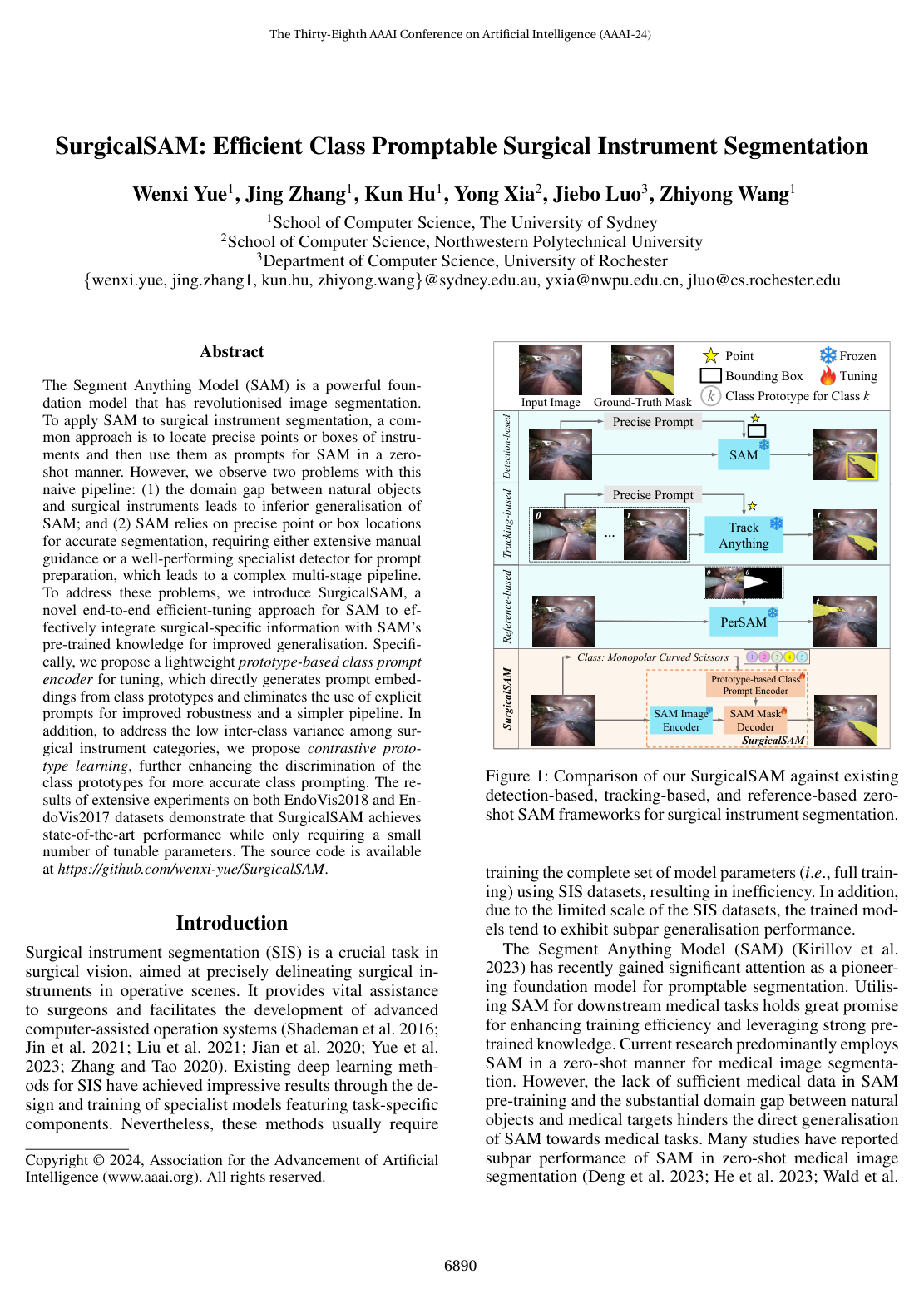
SurgicalSAM: Efficient Class Promptable Surgical Instrument Segmentation
Wenxi Yue, Jing Zhang, Kun Hu, Yong Xia, Jiebo Luo, Zhiyong Wang
Why you should read this
Proposes an end-to-end framework that efficiently adapts the Segment Anything Model for surgical instrument segmentation by replacing fragile manual bounding-box prompts with learned class prototypes and contrastive learning.
The Segment Anything Model (SAM) is a powerful foundation model that has revolutionised image segmentation. To apply SAM to surgical instrument segmentation, a common approach is to locate precise points or boxes of instruments and then use them as prompts for SAM in a zero-shot manner. However, we observe two problems with this naive pipeline: (1) the domain gap between natural objects and surgical instruments leads to inferior generalisation of SAM; and (2) SAM relies on precise point or box locations for accurate segmentation, requiring either extensive manual guidance or a well-performing specialist detector for prompt preparation, which leads to a complex multi-stage pipeline. To address these problems, we introduce SurgicalSAM, a novel end-to-end efficient-tuning approach for SAM to effectively integrate surgical-specific information with SAM's pre-trained knowledge for improved generalisation. Specifically, we propose a lightweight prototype-based class prompt encoder for tuning, which directly generates prompt embeddings from class prototypes and eliminates the use of explicit prompts for improved robustness and a simpler pipeline. In addition, to address the low inter-class variance among surgical instrument categories, we propose contrastive prototype learning, further enhancing the discrimination of the class prototypes for more accurate class prompting. The results of extensive experiments on both EndoVis2018 and EndoVis2017 datasets demonstrate that SurgicalSAM achieves state-of-the-art performance while only requiring a small number of tunable parameters. The source code is available at https://github.com/wenxi-yue/SurgicalSAM.
Added
2026-09-26
Diffusion Adversarial Representation Learning for Self-supervised Vessel Segmentation
Boah Kim, Yujin Oh, Jong Chul Ye
Why you should read this
Proposes a self-supervised diffusion adversarial learning framework that isolates complex background signals to achieve accurate, single-step vessel segmentation across retinal and angiography images without manual annotations.
Vessel segmentation in medical images is one of the important tasks in the diagnosis of vascular diseases and therapy planning. Although learning-based segmentation approaches have been extensively studied, a large amount of ground-truth labels are required in supervised methods and confusing background structures make neural networks hard to segment vessels in an unsupervised manner. To address this, here we introduce a novel diffusion adversarial representation learning (DARL) model that leverages a denoising diffusion probabilistic model with adversarial learning, and apply it to vessel segmentation. In particular, for self-supervised vessel segmentation, DARL learns the background signal using a diffusion module, which lets a generation module effectively provide vessel representations. Also, by adversarial learning based on the proposed switchable spatially-adaptive denormalization, our model estimates synthetic fake vessel images as well as vessel segmentation masks, which further makes the model capture vessel-relevant semantic information. Once the proposed model is trained, the model generates segmentation masks in a single step and can be applied to general vascular structure segmentation of coronary angiography and retinal images. Experimental results on various datasets show that our method significantly outperforms existing unsupervised and self-supervised vessel segmentation methods.
Added
2026-09-26
ResUNet++: An Advanced Architecture for Medical Image Segmentation
Debesh Jha, Pia H. Smedsrud, Michael A. Riegler, Dag Johansen, Thomas de Lange, Pal Halvorsen, Havard D. Johansen
Why you should read this
Introduces ResUNet++, an advanced deep learning architecture that improves pixel-wise polyp segmentation in colonoscopy images and significantly outperforms standard U-Net and ResUNet models on public benchmarks.
Accurate computer-aided polyp detection and segmentation during colonoscopy examinations can help endoscopists resect abnormal tissue and thereby decrease chances of polyps growing into cancer. Towards developing a fully automated model for pixel-wise polyp segmentation, we propose ResUNet++, which is an improved ResUNet architecture for colonoscopic image segmentation. Our experimental evaluations show that the suggested architecture produces good segmentation results on publicly available datasets. Furthermore, ResUNet++ significantly outperforms U-Net and ResUNet, two key state-of-the-art deep learning architectures, by achieving high evaluation scores with a dice coefficient of 81.33%, and a mean Intersection over Union (mIoU) of 79.27% for the Kvasir-SEG dataset and a dice coefficient of 79.55%, and a mIoU of 79.62% with CVC-612 dataset.
Added
2026-09-25
Medical Transformer: Gated Axial-Attention for Medical Image Segmentation
Jeya Maria Jose Valanarasu, Poojan Oza, Ilker Hacihaliloglu, Vishal M. Patel
Why you should read this
Introduces Medical Transformer (MedT), a gated axial-attention architecture paired with a local-global training strategy designed to overcome the data-scarcity bottleneck of vision transformers and outperform convolutional baselines in medical image segmentation.
Over the past decade, Deep Convolutional Neural Networks have been widely adopted for medical image segmentation and shown to achieve adequate performance. However, due to the inherent inductive biases present in the convolutional architectures, they lack understanding of long-range dependencies in the image. Recently proposed Transformer-based architectures that leverage self-attention mechanism encode long-range dependencies and learn representations that are highly expressive. This motivates us to explore Transformer-based solutions and study the feasibility of using Transformer-based network architectures for medical image segmentation tasks. Majority of existing Transformer-based network architectures proposed for vision applications require large-scale datasets to train properly. However, compared to the datasets for vision applications, for medical imaging the number of data samples is relatively low, making it difficult to efficiently train transformers for medical applications. To this end, we propose a Gated Axial-Attention model which extends the existing architectures by introducing an additional control mechanism in the self-attention module. Furthermore, to train the model effectively on medical images, we propose a Local-Global training strategy (LoGo) which further improves the performance. Specifically, we operate on the whole image and patches to learn global and local features, respectively. The proposed Medical Transformer (MedT) is evaluated on three different medical image segmentation datasets and it is shown that it achieves better performance than the convolutional and other related transformer-based architectures. Code: this https URL
Added
2026-09-25
TransFuse: Fusing Transformers and CNNs for Medical Image Segmentation
Yundong Zhang, Huiye Liu, Qiang Hu
Why you should read this
Proposes a parallel Transformer-CNN architecture that captures both global context and fine spatial features through a novel fusion module, setting new benchmarks across 2D and 3D medical image segmentation while cutting model parameters and inference latency.
Medical image segmentation - the prerequisite of numerous clinical needs - has been significantly prospered by recent advances in convolutional neural networks (CNNs). However, it exhibits general limitations on modeling explicit long-range relation, and existing cures, resorting to building deep encoders along with aggressive downsampling operations, leads to redundant deepened networks and loss of localized details. Hence, the segmentation task awaits a better solution to improve the efficiency of modeling global contexts while maintaining a strong grasp of low-level details. In this paper, we propose a novel parallel-in-branch architecture, TransFuse, to address this challenge. TransFuse combines Transformers and CNNs in a parallel style, where both global dependency and low-level spatial details can be efficiently captured in a much shallower manner. Besides, a novel fusion technique - BiFusion module is created to efficiently fuse the multi-level features from both branches. Extensive experiments demonstrate that TransFuse achieves the newest state-of-the-art results on both 2D and 3D medical image sets including polyp, skin lesion, hip, and prostate segmentation, with significant parameter decrease and inference speed improvement.
Added
2026-09-25
CE-Net: Context Encoder Network for 2D Medical Image Segmentation
Zaiwang Gu, Jun Cheng, Huazhu Fu, Kang Zhou, Huaying Hao, Yitian Zhao, Tianyang Zhang, Shenghua Gao, Jiang Liu
Why you should read this
Proposes CE-Net, a medical image segmentation architecture that integrates dense atrous convolution and residual multi-kernel pooling to capture rich contextual features while preserving critical spatial details lost in conventional U-Net pooling.
Medical image segmentation is an important step in medical image analysis. With the rapid development of convolutional neural network in image processing, deep learning has been used for medical image segmentation, such as optic disc segmentation, blood vessel detection, lung segmentation, cell segmentation, etc. Previously, U-net based approaches have been proposed. However, the consecutive pooling and strided convolutional operations lead to the loss of some spatial information. In this paper, we propose a context encoder network (referred to as CE-Net) to capture more high-level information and preserve spatial information for 2D medical image segmentation. CE-Net mainly contains three major components: a feature encoder module, a context extractor and a feature decoder module. We use pretrained ResNet block as the fixed feature extractor. The context extractor module is formed by a newly proposed dense atrous convolution (DAC) block and residual multi-kernel pooling (RMP) block. We applied the proposed CE-Net to different 2D medical image segmentation tasks. Comprehensive results show that the proposed method outperforms the original U-Net method and other state-of-the-art methods for optic disc segmentation, vessel detection, lung segmentation, cell contour segmentation and retinal optical coherence tomography layer segmentation.
Added
2026-09-18
Swin UNETR: Swin Transformers for Semantic Segmentation of Brain Tumors in MRI Images
Ali Hatamizadeh, Vishwesh Nath, Yucheng Tang, Dong Yang, Holger Roth, Daguang Xu
Why you should read this
Presents Swin UNETR, an architecture that couples a hierarchical shifted-window transformer encoder with a convolutional decoder to capture multi-scale contextual dependencies for multi-modal 3D brain tumor MRI segmentation.
Semantic segmentation of brain tumors is a fundamental medical image analysis task involving multiple MRI imaging modalities that can assist clinicians in diagnosing the patient and successively studying the progression of the malignant entity. In recent years, Fully Convolutional Neural Networks (FCNNs) approaches have become the de facto standard for 3D medical image segmentation. The popular "U-shaped" network architecture has achieved state-of-the-art performance benchmarks on different 2D and 3D semantic segmentation tasks and across various imaging modalities. However, due to the limited kernel size of convolution layers in FCNNs, their performance of modeling long-range information is sub-optimal, and this can lead to deficiencies in the segmentation of tumors with variable sizes. On the other hand, transformer models have demonstrated excellent capabilities in capturing such long-range information in multiple domains, including natural language processing and computer vision. Inspired by the success of vision transformers and their variants, we propose a novel segmentation model termed Swin UNEt TRansformers (Swin UNETR). Specifically, the task of 3D brain tumor semantic segmentation is reformulated as a sequence to sequence prediction problem wherein multi-modal input data is projected into a 1D sequence of embedding and used as an input to a hierarchical Swin transformer as the encoder. The swin transformer encoder extracts features at five different resolutions by utilizing shifted windows for computing self-attention and is connected to an FCNN-based decoder at each resolution via skip connections. We have participated in BraTS 2021 segmentation challenge, and our proposed model ranks among the top-performing approaches in the validation phase. Code: this https URL
Added
2026-09-16
Swin-Unet: Unet-like Pure Transformer for Medical Image Segmentation
Hu Cao, Yueyue Wang, Joy Chen, Dongsheng Jiang, Xiaopeng Zhang, Qi Tian, Manning Wang
Why you should read this
Introduces Swin-Unet, a pure Transformer U-shaped encoder-decoder network with skip connections that learns long-range contextual interactions to outperform conventional convolutional and hybrid baselines on multi-organ and cardiac image segmentation.
In the past few years, convolutional neural networks (CNNs) have achieved milestones in medical image analysis. Especially, the deep neural networks based on U-shaped architecture and skip-connections have been widely applied in a variety of medical image tasks. However, although CNN has achieved excellent performance, it cannot learn global and long-range semantic information interaction well due to the locality of the convolution operation. In this paper, we propose Swin-Unet, which is an Unet-like pure Transformer for medical image segmentation. The tokenized image patches are fed into the Transformer-based U-shaped Encoder-Decoder architecture with skip-connections for local-global semantic feature learning. Specifically, we use hierarchical Swin Transformer with shifted windows as the encoder to extract context features. And a symmetric Swin Transformer-based decoder with patch expanding layer is designed to perform the up-sampling operation to restore the spatial resolution of the feature maps. Under the direct down-sampling and up-sampling of the inputs and outputs by 4x, experiments on multi-organ and cardiac segmentation tasks demonstrate that the pure Transformer-based U-shaped Encoder-Decoder network outperforms those methods with full-convolution or the combination of transformer and convolution. The codes and trained models will be publicly available at this https URL.
Added
2026-09-14
Generalised Dice Overlap as a Deep Learning Loss Function for Highly Unbalanced Segmentations
Carole H. Sudre, Wenqi Li, Tom Kamiel Magda Vercauteren, Sébastien Ourselin, M. Jorge Cardoso
Why you should read this
Proposes adapting the Generalized Dice overlap into a deep learning loss function to solve severe class imbalance problems in 2D and 3D medical image segmentation.
Deep-learning has proved in recent years to be a powerful tool for image analysis and is now widely used to segment both 2D and 3D medical images. Deep-learning segmentation frameworks rely not only on the choice of network architecture but also on the choice of loss function. When the segmentation process targets rare observations, a severe class imbalance is likely to occur between candidate labels, thus resulting in sub-optimal performance. In order to mitigate this issue, strategies such as the weighted cross-entropy function, the sensitivity function or the Dice loss function, have been proposed. In this work, we investigate the behavior of these loss functions and their sensitivity to learning rate tuning in the presence of different rates of label imbalance across 2D and 3D segmentation tasks. We also propose to use the class re-balancing properties of the Generalized Dice overlap, a known metric for segmentation assessment, as a robust and accurate deep-learning loss function for unbalanced tasks.
Added
2026-09-14
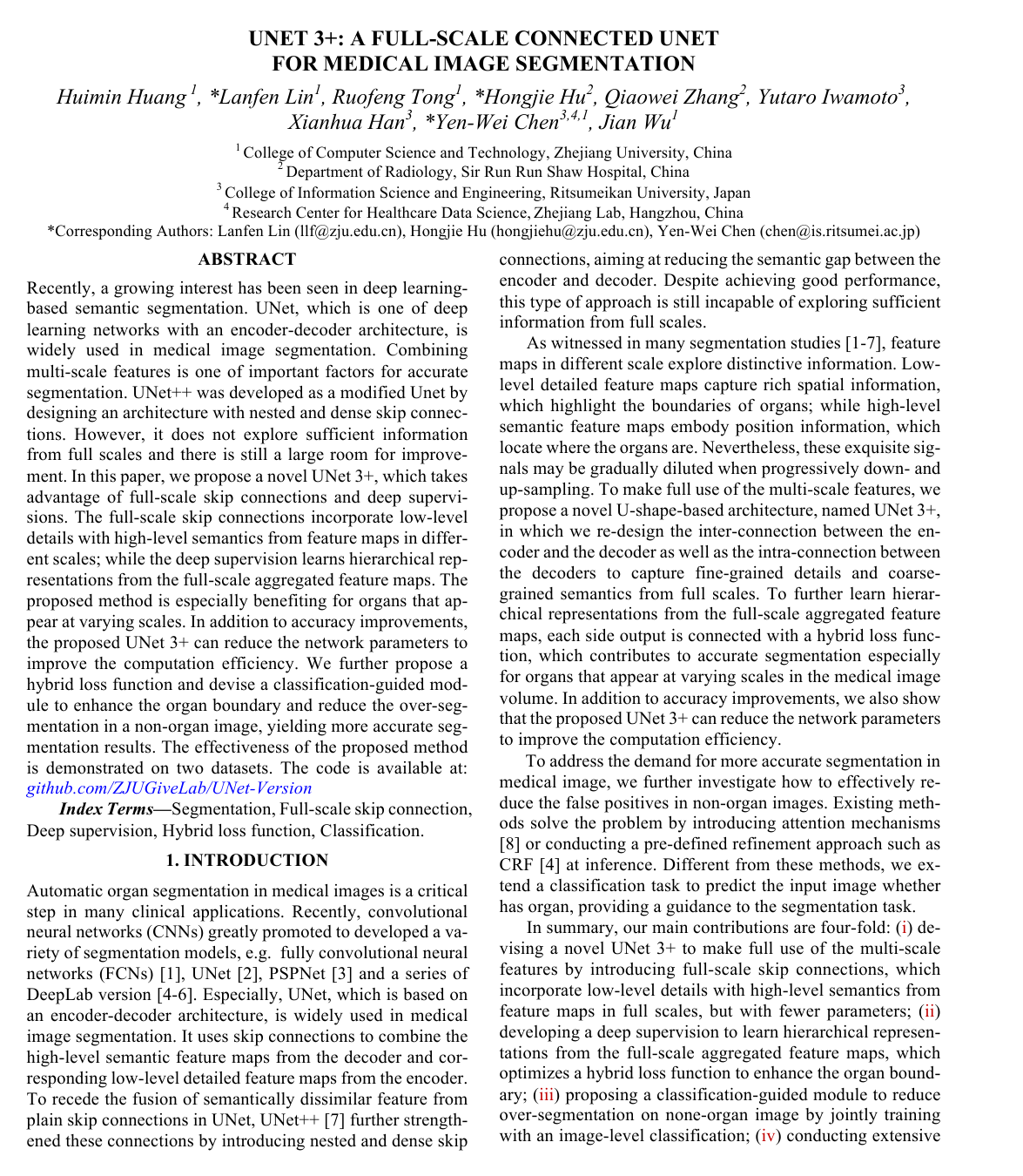
UNet 3+: A Full-Scale Connected UNet for Medical Image Segmentation
Huimin Huang, Lanfen Lin, Ruofeng Tong, Hongjie Hu, Qiaowei Zhang, Yutaro Iwamoto, Xianhua Han, Yen-Wei Chen, Jian Wu
Why you should read this
Introduces UNet 3+, a medical image segmentation architecture that combines full-scale skip connections with deep supervision to capture multi-scale anatomical structures with higher accuracy and fewer parameters than previous UNet variants.
Recently, a growing interest has been seen in deep learning-based semantic segmentation. UNet, which is one of deep learning networks with an encoder-decoder architecture, is widely used in medical image segmentation. Combining multi-scale features is one of important factors for accurate segmentation. UNet++ was developed as a modified Unet by designing an architecture with nested and dense skip connections. However, it does not explore sufficient information from full scales and there is still a large room for improvement. In this paper, we propose a novel UNet 3+, which takes advantage of full-scale skip connections and deep supervisions. The full-scale skip connections incorporate low-level details with high-level semantics from feature maps in different scales; while the deep supervision learns hierarchical representations from the full-scale aggregated feature maps. The proposed method is especially benefiting for organs that appear at varying scales. In addition to accuracy improvements, the proposed UNet 3+ can reduce the network parameters to improve the computation efficiency. We further propose a hybrid loss function and devise a classification-guided module to enhance the organ boundary and reduce the over-segmentation in a non-organ image, yielding more accurate segmentation results. The effectiveness of the proposed method is demonstrated on two datasets. The code is available at: this http URL
Added
2026-09-14
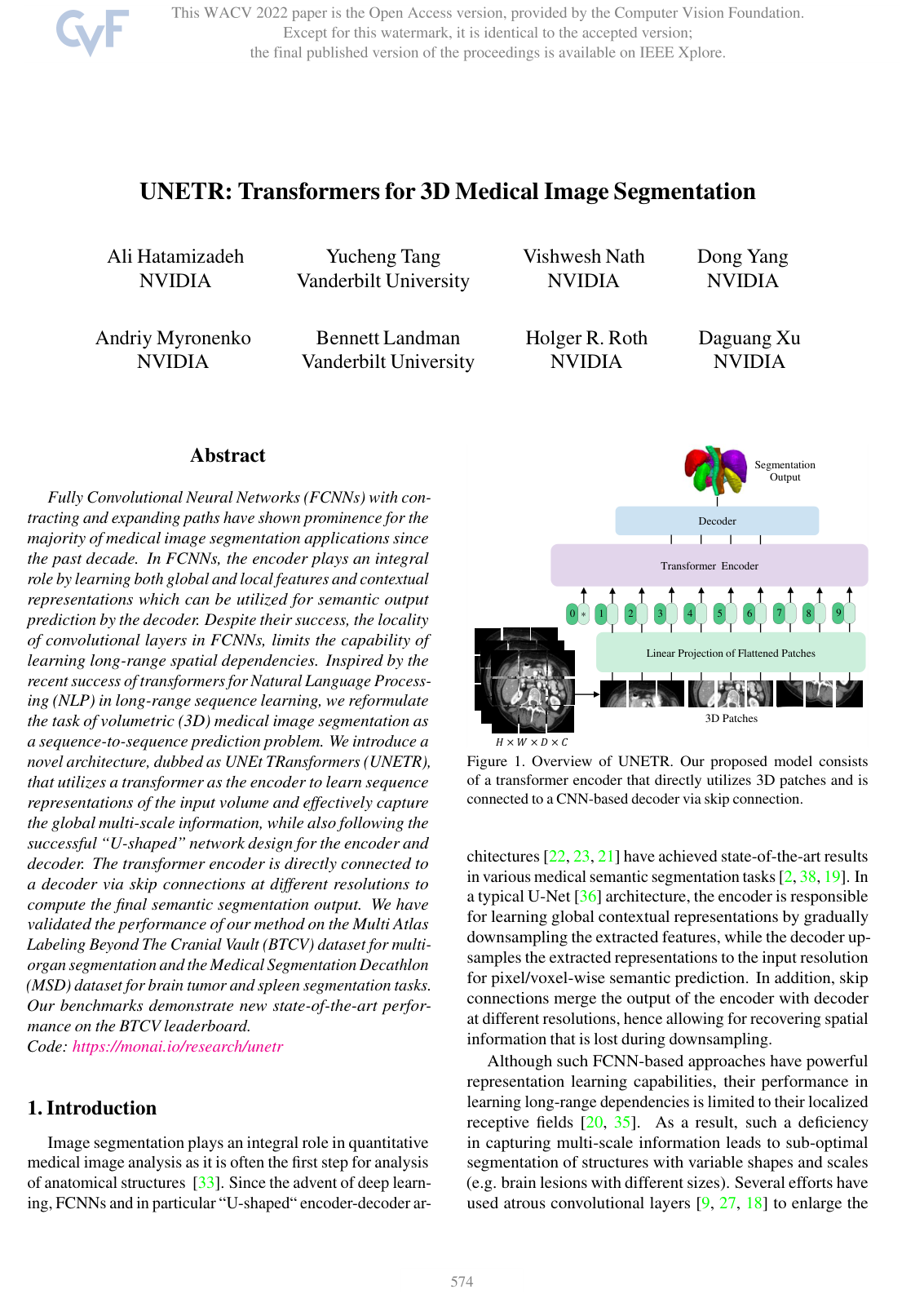
UNETR: Transformers for 3D Medical Image Segmentation
Ali Hatamizadeh, Dong Yang, Holger R. Roth, Daguang Xu
Why you should read this
Introduces UNETR, a volumetric medical image segmentation architecture that connects a pure vision transformer encoder to a CNN decoder via multi-scale skip connections, achieving state-of-the-art performance on 3D CT and MRI benchmarks by capturing global spatial dependencies.
Fully Convolutional Neural Networks (FCNNs) with con-tracting and expanding paths have shown prominence for the majority of medical image segmentation applications since the past decade. In FCNNs, the encoder plays an integral role by learning both global and local features and contextual representations which can be utilized for semantic output prediction by the decoder. Despite their success, the locality of convolutional layers in FCNNs, limits the capability of learning long-range spatial dependencies. Inspired by the recent success of transformers for Natural Language Process-ing (NLP) in long-range sequence learning, we reformulate the task of volumetric (3D) medical image segmentation as a sequence-to-sequence prediction problem. We introduce a novel architecture, dubbed as UNEt TRansformers (UNETR), that utilizes a transformer as the encoder to learn sequence representations of the input volume and effectively capture the global multi-scale information, while also following the successful “U-shaped” network design for the encoder and decoder. The transformer encoder is directly connected to a decoder via skip connections at different resolutions to compute the final semantic segmentation output. We have validated the performance of our method on the Multi Atlas Labeling Beyond The Cranial Vault (BTCV) dataset for multi-organ segmentation and the Medical Segmentation Decathlon (MSD) dataset for brain tumor and spleen segmentation tasks. Our benchmarks demonstrate new state-of-the-art perfor-mance on the BTCV leaderboard. Code: https://monai.io/research/unetr
Added
2026-09-14
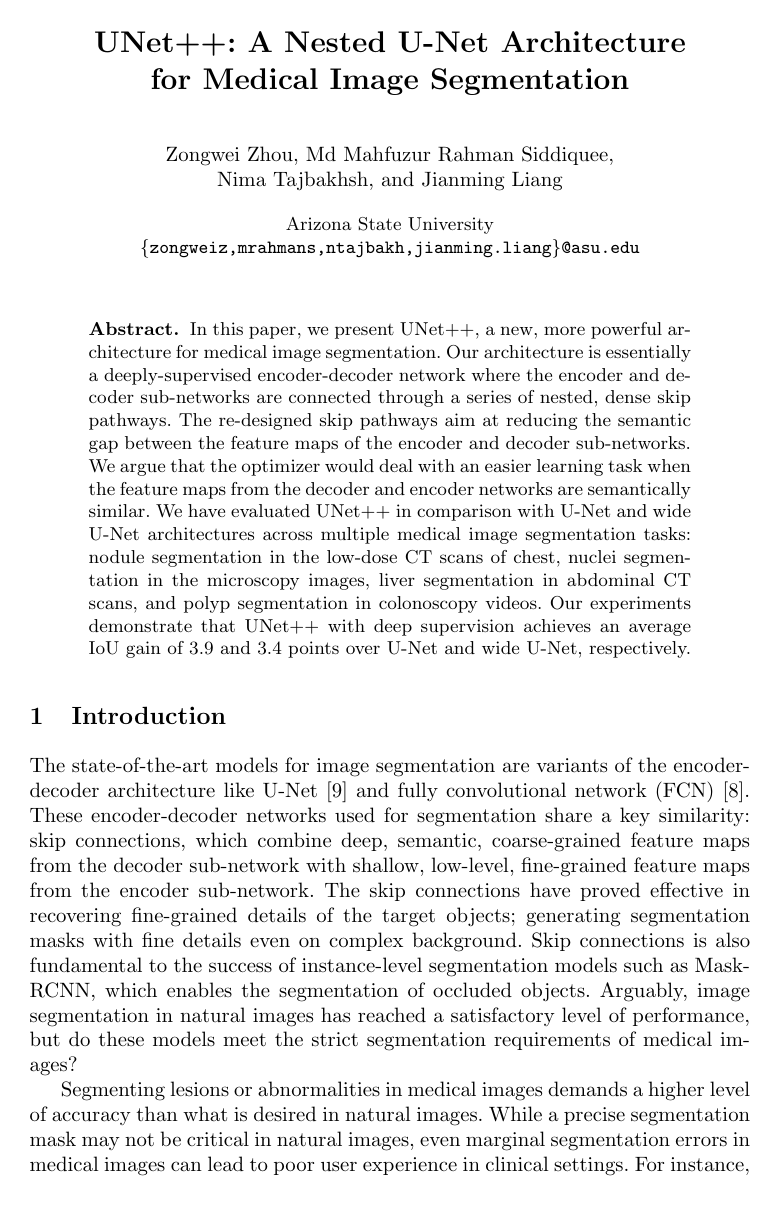
UNet++: A Nested U-Net Architecture for Medical Image Segmentation
Zongwei Zhou, Md Mahfuzur Rahman Siddiquee, Nima Tajbakhsh, Jianming Liang
Why you should read this
Proposes UNet++, a deeply-supervised encoder-decoder architecture with nested, dense skip pathways that reduces the semantic gap between feature representations to achieve superior segmentation performance across diverse medical imaging modalities.
In this paper, we present UNet++, a new, more powerful architecture for medical image segmentation. Our architecture is essentially a deeply-supervised encoder-decoder network where the encoder and decoder sub-networks are connected through a series of nested, dense skip pathways. The re-designed skip pathways aim at reducing the semantic gap between the feature maps of the encoder and decoder sub-networks. We argue that the optimizer would deal with an easier learning task when the feature maps from the decoder and encoder networks are semantically similar. We have evaluated UNet++ in comparison with U-Net and wide U-Net architectures across multiple medical image segmentation tasks: nodule segmentation in the low-dose CT scans of chest, nuclei segmentation in the microscopy images, liver segmentation in abdominal CT scans, and polyp segmentation in colonoscopy videos. Our experiments demonstrate that UNet++ with deep supervision achieves an average IoU gain of 3.9 and 3.4 points over U-Net and wide U-Net, respectively.
Added
2026-09-07