
keyword
medical image analysis
Medical image analysis is an interdisciplinary field that applies computational methods, computer vision, and machine learning to process, interpret, and extract meaningful quantitative information from biomedical images. Operating on visual data acquired through modalities such as magnetic resonance imaging, computed tomography, ultrasound, X-rays, and digital pathology, the discipline encompasses key tasks including image segmentation, anomaly detection, tissue classification, and spatial registration. By automating feature extraction and identifying subtle physiological or pathological patterns, medical image analysis assists healthcare professionals in disease screening, clinical diagnosis, treatment planning, and therapeutic monitoring.
6 items
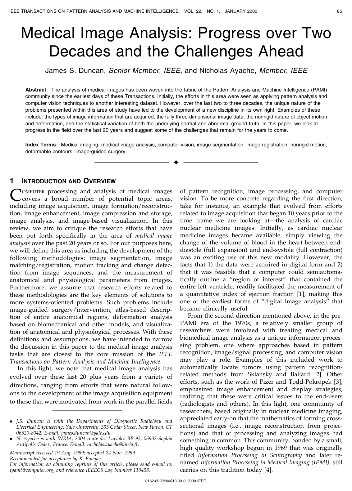
Medical Image Analysis: Progress over Two Decades and the Challenges Ahead
J. Duncan, N. Ayache
Why you should read this
Synthesizes twenty years of foundational advances in medical image analysis across segmentation, registration, motion tracking, and image-guided surgery while defining critical open problems for integrating biomechanical models and physiological data.
Added
2026-09-25
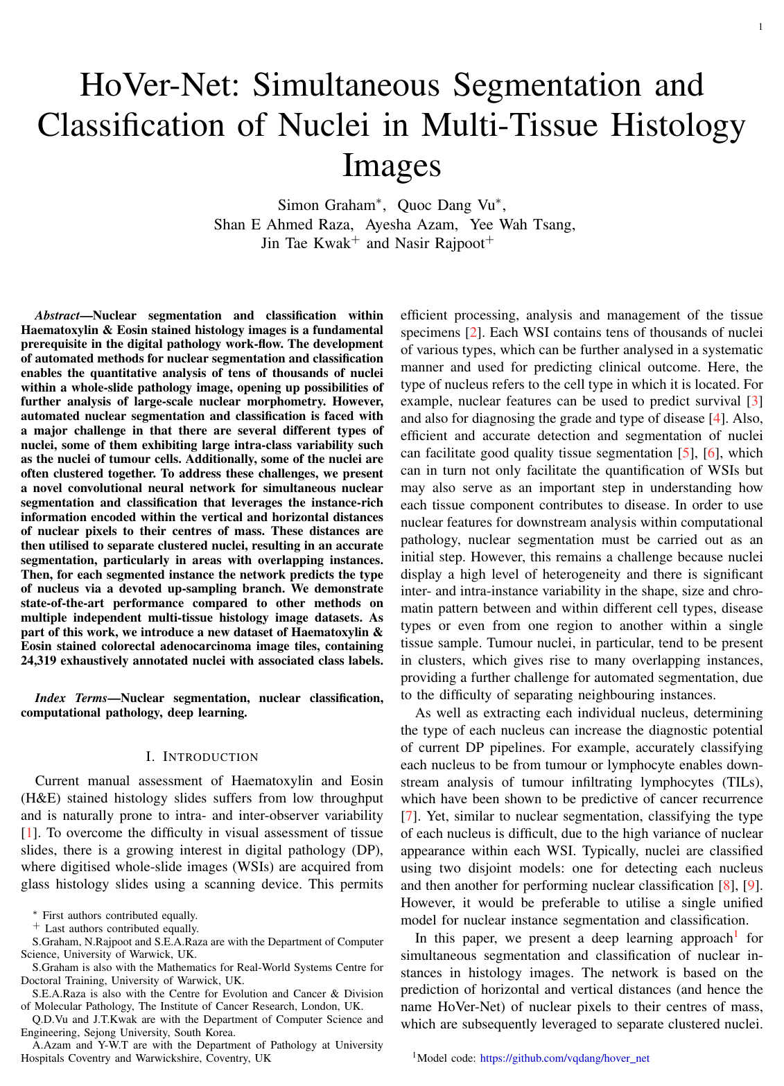
Hover-Net: Simultaneous segmentation and classification of nuclei in multi-tissue histology images
Simon Graham, Quoc Dang Vu, Shan E Ahmed Raza, Ayesha Azam, Yee Wah Tsang, Jin Tae Kwak, Nasir Rajpoot
Why you should read this
Presents HoVer-Net, a deep learning architecture that uses horizontal and vertical pixel distance maps to accurately separate clustered nuclei and simultaneously classify cell types across multi-tissue histology images.
Nuclear segmentation and classification within Haematoxylin & Eosin stained histology images is a fundamental prerequisite in the digital pathology work-flow. The development of automated methods for nuclear segmentation and classification enables the quantitative analysis of tens of thousands of nuclei within a whole-slide pathology image, opening up possibilities of further analysis of large-scale nuclear morphometry. However, automated nuclear segmentation and classification is faced with a major challenge in that there are several different types of nuclei, some of them exhibiting large intra-class variability such as the tumour cells. Additionally, some of the nuclei are often clustered together. To address these challenges, we present a novel convolutional neural network for simultaneous nuclear segmentation and classification that leverages the instance-rich information encoded within the vertical and horizontal distances of nuclear pixels to their centres of mass. These distances are then utilised to separate clustered nuclei, resulting in an accurate segmentation, particularly in areas with overlapping instances. Then for each segmented instance, the network predicts the type of nucleus via a devoted up-sampling branch. We demonstrate state-of-the-art performance compared to other methods on multiple independent multi-tissue histology image datasets. As part of this work, we introduce a new dataset of Haematoxylin & Eosin stained colorectal adenocarcinoma image tiles, containing 24,319 exhaustively annotated nuclei with associated class labels.
Added
2026-09-25
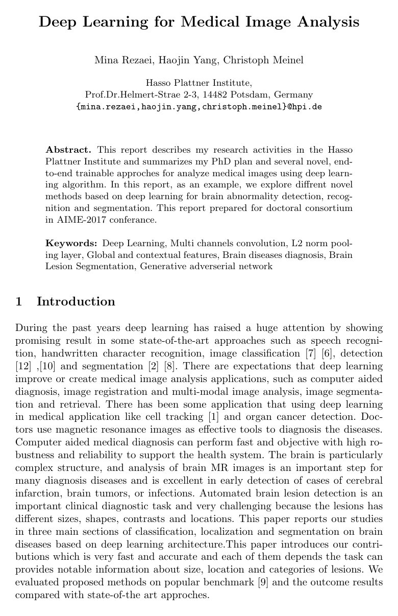
Deep Learning for Medical Image Analysis
Mina Rezaei, Haojin Yang, Christoph Meinel
Why you should read this
Proposes end-to-end deep learning methods for automated brain abnormality detection, recognition, and segmentation to advance clinical neuroimaging analysis.
This report describes my research activities in the Hasso Plattner Institute and summarizes my Ph.D. plan and several novels, end-to-end trainable approaches for analyzing medical images using deep learning algorithm. In this report, as an example, we explore different novel methods based on deep learning for brain abnormality detection, recognition, and segmentation. This report prepared for the doctoral consortium in the AIME-2017 conference.
Added
2026-09-14
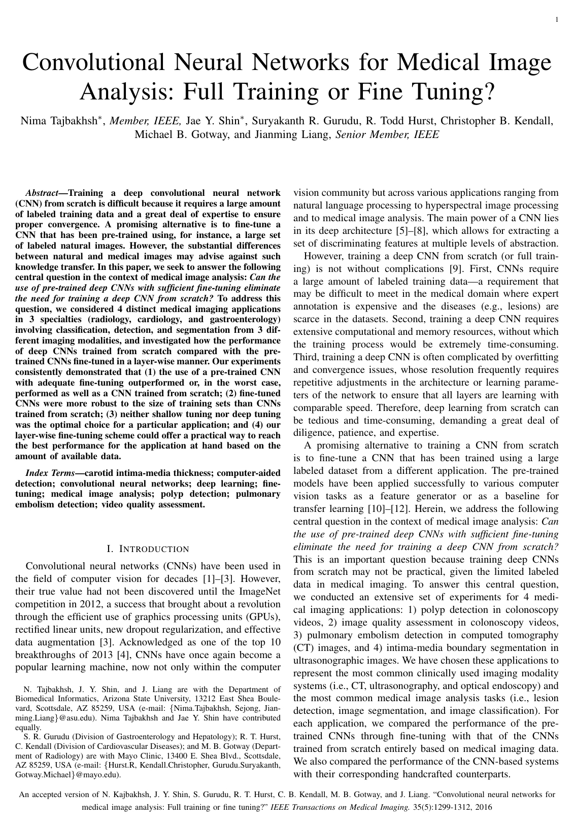
Convolutional Neural Networks for Medical Image Analysis: Full Training or Fine Tuning?
Nima Tajbakhsh, Jae Y. Shin, Suryakanth R. Gurudu, R. Todd Hurst, Christopher B. Kendall, Michael B. Gotway, Jianming Liang
Why you should read this
Establishes that fine-tuning pre-trained convolutional neural networks consistently matches or exceeds the performance of models trained from scratch across diverse medical imaging tasks, providing a practical layer-wise strategy to overcome scarce clinical training data.
Training a deep convolutional neural network (CNN) from scratch is difficult because it requires a large amount of labeled training data and a great deal of expertise to ensure proper convergence. A promising alternative is to fine-tune a CNN that has been pre-trained using, for instance, a large set of labeled natural images. However, the substantial differences between natural and medical images may advise against such knowledge transfer. In this paper, we seek to answer the following central question in the context of medical image analysis: \emph{Can the use of pre-trained deep CNNs with sufficient fine-tuning eliminate the need for training a deep CNN from scratch?} To address this question, we considered 4 distinct medical imaging applications in 3 specialties (radiology, cardiology, and gastroenterology) involving classification, detection, and segmentation from 3 different imaging modalities, and investigated how the performance of deep CNNs trained from scratch compared with the pre-trained CNNs fine-tuned in a layer-wise manner. Our experiments consistently demonstrated that (1) the use of a pre-trained CNN with adequate fine-tuning outperformed or, in the worst case, performed as well as a CNN trained from scratch; (2) fine-tuned CNNs were more robust to the size of training sets than CNNs trained from scratch; (3) neither shallow tuning nor deep tuning was the optimal choice for a particular application; and (4) our layer-wise fine-tuning scheme could offer a practical way to reach the best performance for the application at hand based on the amount of available data.
Added
2026-09-13
A survey on deep learning in medical image analysis
Geert Litjens, Thijs Kooi, Babak Ehteshami Bejnordi, Arnaud Arindra Adiyoso Setio, Francesco Ciompi, Mohsen Ghafoorian, Jeroen A. W. M. van der Laak, Bram van Ginneken, Clara I. Sánchez
Why you should read this
Synthesizes over 300 foundational studies across major clinical domains to map deep learning architectures directly to essential tasks like lesion detection, anatomical segmentation, and image registration while identifying key technical bottlenecks.
Deep learning algorithms, in particular convolutional networks, have rapidly become a methodology of choice for analyzing medical images. This paper reviews the major deep learning concepts pertinent to medical image analysis and summarizes over 300 contributions to the field, most of which appeared in the last year. We survey the use of deep learning for image classification, object detection, segmentation, registration, and other tasks and provide concise overviews of studies per application area. Open challenges and directions for future research are discussed.
Added
2026-09-13
Brain tumor segmentation with Deep Neural Networks
Mohammad Havaei, Axel Davy, David Warde-Farley, Antoine Biard, Aaron Courville, Yoshua Bengio, Chris Pal, Pierre-Marc Jodoin, Hugo Larochelle
Why you should read this
Proposes a multi-path convolutional neural network for MRI brain tumor segmentation that combines local and global context alongside a two-phase training strategy to handle label imbalance, outperforming prior methods on the BRATS benchmark while running over thirty times faster.
In this paper, we present a fully automatic brain tumor segmentation method based on Deep Neural Networks (DNNs). The proposed networks are tailored to glioblastomas (both low and high grade) pictured in MR images. By their very nature, these tumors can appear anywhere in the brain and have almost any kind of shape, size, and contrast. These reasons motivate our exploration of a machine learning solution that exploits a flexible, high capacity DNN while being extremely efficient. Here, we give a description of different model choices that we've found to be necessary for obtaining competitive performance. We explore in particular different architectures based on Convolutional Neural Networks (CNN), i.e. DNNs specifically adapted to image data. We present a novel CNN architecture which differs from those traditionally used in computer vision. Our CNN exploits both local features as well as more global contextual features simultaneously. Also, different from most traditional uses of CNNs, our networks use a final layer that is a convolutional implementation of a fully connected layer which allows a 40 fold speed up. We also describe a 2-phase training procedure that allows us to tackle difficulties related to the imbalance of tumor labels. Finally, we explore a cascade architecture in which the output of a basic CNN is treated as an additional source of information for a subsequent CNN. Results reported on the 2013 BRATS test dataset reveal that our architecture improves over the currently published state-of-the-art while being over 30 times faster.
Added
2026-09-12