
keyword
computational pathology
Computational pathology is an interdisciplinary field that applies artificial intelligence, computer vision, and advanced data science methods to analyze digitized biological tissue samples and whole-slide microscopic images. It focuses on transforming high-resolution histology slides into quantitative, interpretable data to assist pathologists and researchers in disease diagnosis, cancer subtyping, prognosis, and therapeutic response prediction. By automating complex visual tasks such as cell and nuclear segmentation, tissue structure classification, and morphological feature extraction, computational pathology enhances diagnostic accuracy, consistency, and efficiency across clinical workflows and biomedical research.
4 items
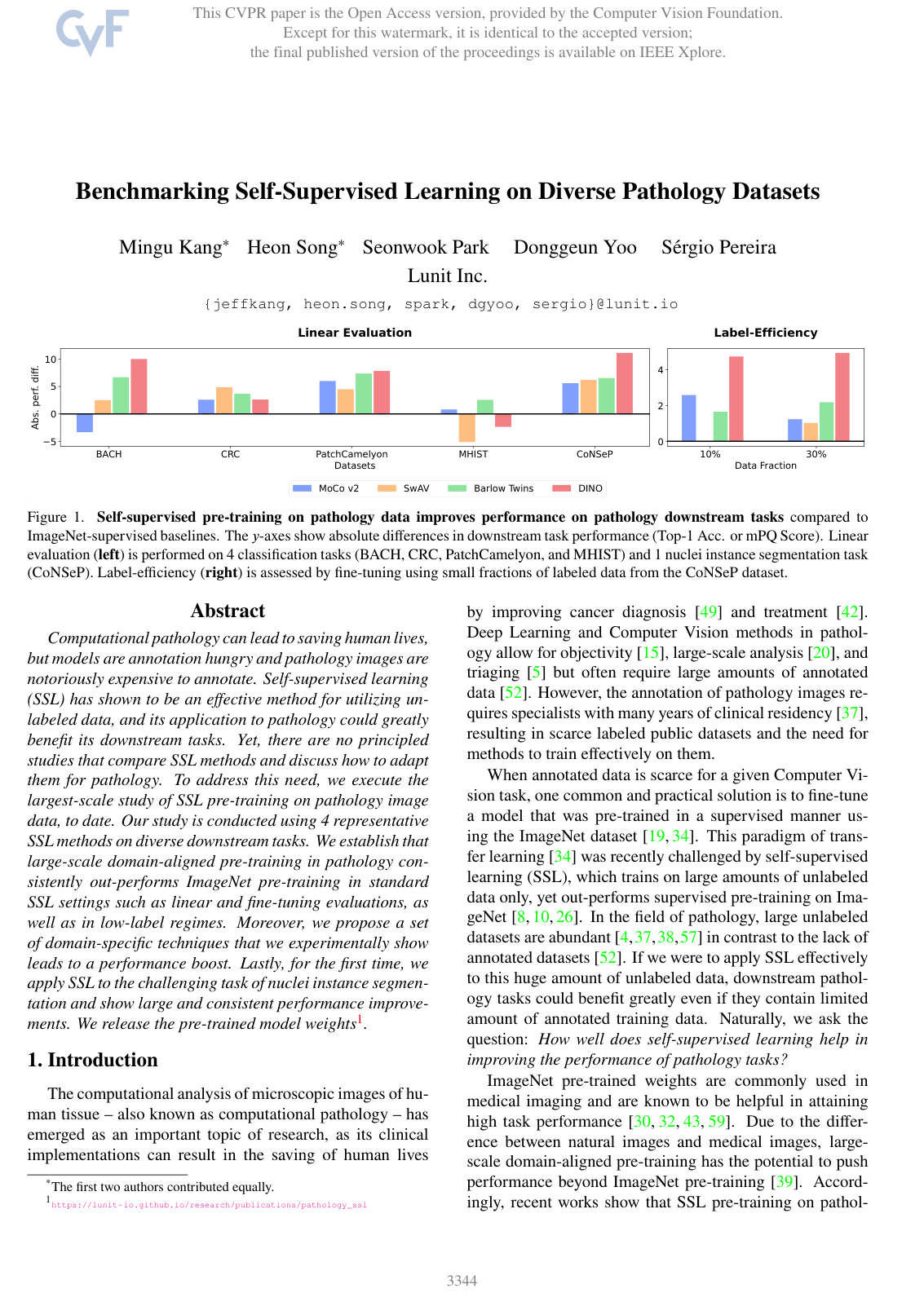
Benchmarking Self-Supervised Learning on Diverse Pathology Datasets
Mingu Kang, Heon Song, Seonwook Park, Donggeun Yoo, Sérgio Pereira
Why you should read this
Presents a large-scale benchmark of self-supervised learning methods on 19 million pathology image patches, demonstrating that domain-aligned pre-training with tailored augmentations consistently outperforms standard ImageNet pre-training across diverse classification and nuclei segmentation tasks, especially in label-scarce settings.
Computational pathology can lead to saving human lives, but models are annotation hungry and pathology images are notoriously expensive to annotate. Self-supervised learning (SSL) has shown to be an effective method for utilizing unlabeled data, and its application to pathology could greatly benefit its downstream tasks. Yet, there are no principled studies that compare SSL methods and discuss how to adapt them for pathology. To address this need, we execute the largest-scale study of SSL pre-training on pathology image data, to date. Our study is conducted using 4 representative SSL methods on diverse downstream tasks. We establish that large-scale domain-aligned pre-training in pathology consistently out-performs ImageNet pre-training in standard SSL settings such as linear and fine-tuning evaluations, as well as in low-label regimes. Moreover, we propose a set of domain-specific techniques that we experimentally show leads to a performance boost. Lastly, for the first time, we apply SSL to the challenging task of nuclei instance segmentation and show large and consistent performance improvements. We release the pre-trained model weights1.
Added
2026-09-26
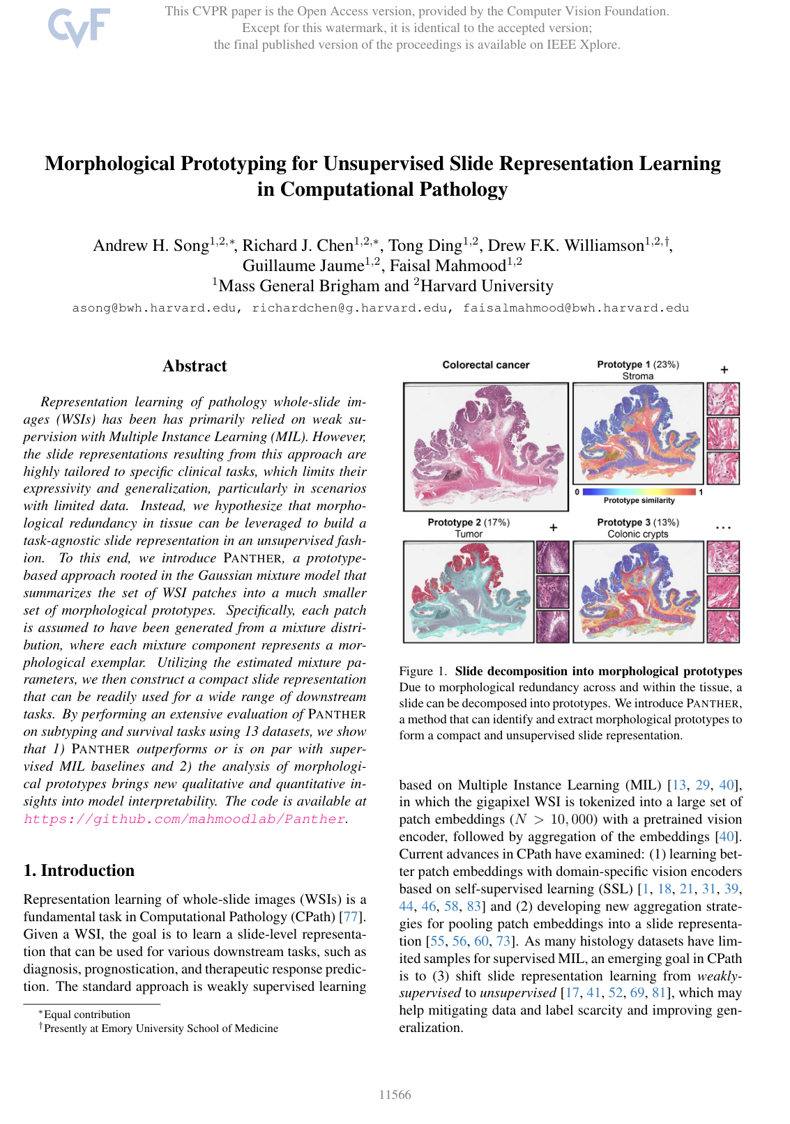
Morphological Prototyping for Unsupervised Slide Representation Learning in Computational Pathology
Andrew H. Song, Richard J. Chen, Tong Ding, Drew F. K. Williamson, Guillaume Jaume, Faisal Mahmood
Why you should read this
Proposes an unsupervised Gaussian mixture model framework that condenses whole-slide images into compact morphological prototypes, matching or outperforming supervised multiple instance learning baselines across subtyping and survival prediction benchmarks while enabling interpretable slide-level analysis.
Representation learning of pathology whole-slide images (WSIs) has been has primarily relied on weak supervision with Multiple Instance Learning (MIL). However, the slide representations resulting from this approach are highly tailored to specific clinical tasks, which limits their expressivity and generalization, particularly in scenarios with limited data. Instead, we hypothesize that morphological redundancy in tissue can be leveraged to build a task-agnostic slide representation in an unsupervised fashion. To this end, we introduce PANTHER, a prototype-based approach rooted in the Gaussian mixture model that summarizes the set of WSI patches into a much smaller set of morphological prototypes. Specifically, each patch is assumed to have been generated from a mixture distribution, where each mixture component represents a morphological exemplar. Utilizing the estimated mixture parameters, we then construct a compact slide representation that can be readily used for a wide range of downstream tasks. By performing an extensive evaluation of PANTHER on subtyping and survival tasks using 13 datasets, we show that 1) PANTHER outperforms or is on par with supervised MIL baselines and 2) the analysis of morphological prototypes brings new qualitative and quantitative insights into model interpretability. The code is available at https://github.com/mahmoodlab/Panther.
Added
2026-09-26
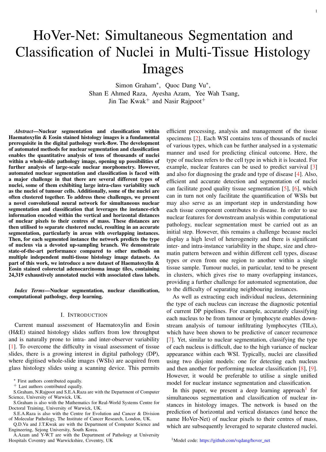
Hover-Net: Simultaneous segmentation and classification of nuclei in multi-tissue histology images
Simon Graham, Quoc Dang Vu, Shan E Ahmed Raza, Ayesha Azam, Yee Wah Tsang, Jin Tae Kwak, Nasir Rajpoot
Why you should read this
Presents HoVer-Net, a deep learning architecture that uses horizontal and vertical pixel distance maps to accurately separate clustered nuclei and simultaneously classify cell types across multi-tissue histology images.
Nuclear segmentation and classification within Haematoxylin & Eosin stained histology images is a fundamental prerequisite in the digital pathology work-flow. The development of automated methods for nuclear segmentation and classification enables the quantitative analysis of tens of thousands of nuclei within a whole-slide pathology image, opening up possibilities of further analysis of large-scale nuclear morphometry. However, automated nuclear segmentation and classification is faced with a major challenge in that there are several different types of nuclei, some of them exhibiting large intra-class variability such as the tumour cells. Additionally, some of the nuclei are often clustered together. To address these challenges, we present a novel convolutional neural network for simultaneous nuclear segmentation and classification that leverages the instance-rich information encoded within the vertical and horizontal distances of nuclear pixels to their centres of mass. These distances are then utilised to separate clustered nuclei, resulting in an accurate segmentation, particularly in areas with overlapping instances. Then for each segmented instance, the network predicts the type of nucleus via a devoted up-sampling branch. We demonstrate state-of-the-art performance compared to other methods on multiple independent multi-tissue histology image datasets. As part of this work, we introduce a new dataset of Haematoxylin & Eosin stained colorectal adenocarcinoma image tiles, containing 24,319 exhaustively annotated nuclei with associated class labels.
Added
2026-09-25
Data-efficient and weakly supervised computational pathology on whole-slide images
Ming Y. Lu, Drew F. K. Williamson, Tiffany Y. Chen, Richard J. Chen, Matteo Barbieri, Faisal Mahmood
Why you should read this
Presents CLAM, a data-efficient deep learning framework that uses clustering-constrained attention to classify gigapixel whole slide images and identify clinically relevant morphological patterns using only slide-level labels.
The rapidly emerging field of computational pathology has the potential to enable objective diagnosis, therapeutic response prediction and identification of new morphological features of clinical relevance. However, deep learning-based computational pathology approaches either require manual annotation of gigapixel whole slide images (WSIs) in fully-supervised settings or thousands of WSIs with slide-level labels in a weakly-supervised setting. Moreover, whole slide level computational pathology methods also suffer from domain adaptation and interpretability issues. These challenges have prevented the broad adaptation of computational pathology for clinical and research purposes. Here we present CLAM - Clustering-constrained attention multiple instance learning, an easy-to-use, high-throughput, and interpretable WSI-level processing and learning method that only requires slide-level labels while being data efficient, adaptable and capable of handling multi-class subtyping problems. CLAM is a deep-learning-based weakly-supervised method that uses attention-based learning to automatically identify sub-regions of high diagnostic value in order to accurately classify the whole slide, while also utilizing instance-level clustering over the representative regions identified to constrain and refine the feature space. In three separate analyses, we demonstrate the data efficiency and adaptability of CLAM and its superior performance over standard weakly-supervised classification. We demonstrate that CLAM models are interpretable and can be used to identify well-known and new morphological features. We further show that models trained using CLAM are adaptable to independent test cohorts, cell phone microscopy images, and biopsies. CLAM is a general-purpose and adaptable method that can be used for a variety of different computational pathology tasks in both clinical and research settings.
Added
2026-09-15